tree in bud opacities in lungs
Citation DOI article data. In radiology the tree-in-bud sign is a finding on a CT scan that indicates some degree of airway obstruction.
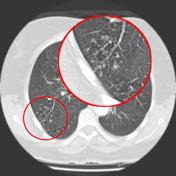
Tree In Bud Sign Lung Radiology Reference Article Radiopaedia Org
Tree-in-bud opacities appear as tiny centrilobular branching structures on CT most often in the lung periphery which resemble budding trees Figure 18-4.
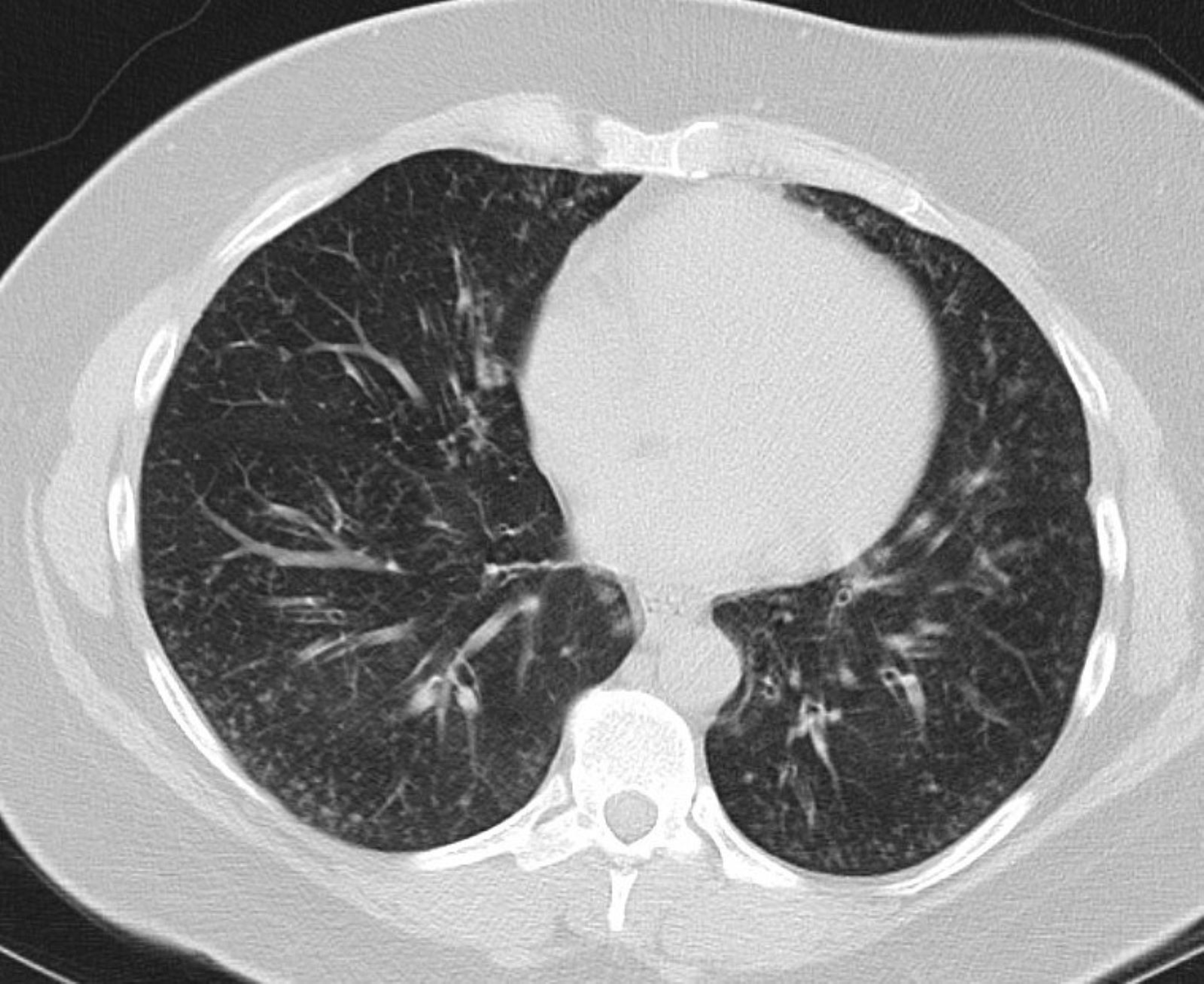
. These opacities are often seen on chest x-rays and are. B Computed tomography in. What causes tree-in-bud opacities.
The tree-in-bud pattern is commonly seen at thin-section computed tomography CT of the lungs. Tree-in-bud sign or pattern describes the CT appearance of multiple areas of centrilobular nodules with a linear branching pattern. Simply put the tree-in-bud pattern can be seen with two main sites of disease 3.
Multiple causes for tree-in-bud TIB opacities an imaging pattern usually seen on chest CT have been reported. Computed tomographic images of the lungs. Distal pulmonary vasculature More specifically the pattern can be manifest because of the following disease processes often in combination.
Opportunistic lung infection in a 73-year-old female patient with selective IgG3 deficit. Bronchial disorders CT lung. The list of the most frequent differential diagnoses for tree-in-bud sign includes infections with Mycobacterium.
The tree-in-bud sign is a nonspecific imaging finding that implies impaction within bronchioles the smallest airway passages in the. Bronchial disorders CT lung. Distal airways more common 2.
It consists of small centrilobular nodules of soft-tissue attenuation connected to. The tree-in-bud sign is a common finding in HRCT scans. These nodules are centered within the secondary pulmonary lobule without involvement of the subpleural lung compatible with a centrilobular distribution.
Note the peripheral tree-in-bud opacities. Tree-in-bud TIB opacities are small round densities in the lung that appear as if a small tree were growing inside the lung. Respiratory infections with mycobacteria bacteria viruses and multiple organisms were the most common.
HRCT image on the axial plane depicts bronchiectasis associated with peribronchial alveolar. 1 refers to a pattern seen on thin-section chest CT in which centrilobular bronchial dilatation and filling by mucus pus or fluid. These are due to filling of the.
Bronchiolesfilled with pus or infla See more. The tree-in-bud sign is a nonspecific imaging finding that implies impaction within. A Computed tomography at admission shows notable infiltrative shadows along the airway in both lung fields.
1 refers to a pattern seen on thin-section chest CT in which centrilobular bronchial dilatation and filling by mucus pus or fluid. Aspiration was the cause in 42 of the 166.

Tree In Bud Pattern At Thin Section Ct Of The Lungs Radiologic Pathologic Overview Radiographics

Kjr Korean Journal Of Radiology
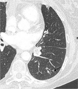
The Tree In Bud Pattern On Chest Ct Radiologic And Microbiologic Correlation Springerlink

Computed Tomography Revealing Tree In Bud Opacities Predominantly In Download Scientific Diagram

Tree In Bud Pattern Pulmonary Tb Eurorad

Chest Ct In Covid 19 What The Radiologist Needs To Know Abstract Europe Pmc

Diffuse Parenchymal Lung Disease And Its Mimics Thoracic Key

Ct Scan Of Chest Revealing Scattered Tree In Bud Opacities In Both Download Scientific Diagram

Tree In Bud Sign And Bronchiectasis Radiology Case Radiopaedia Org

Signs And Patterns Of Lung Disease Chest Radiology The Essentials 2nd Edition
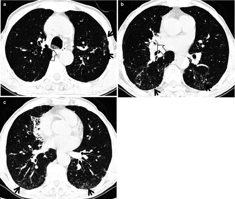
Tree In Bud Sign Radiology Key
Mortality In Rheumatoid Arthritis Patients With Pulmonary Nontuberculous Mycobacterial Disease A Retrospective Cohort Study Plos One

Tree In Bud Pattern At Thin Section Ct Of The Lungs Radiologic Pathologic Overview Radiographics

Causes And Imaging Patterns Of Tree In Bud Opacities Chest
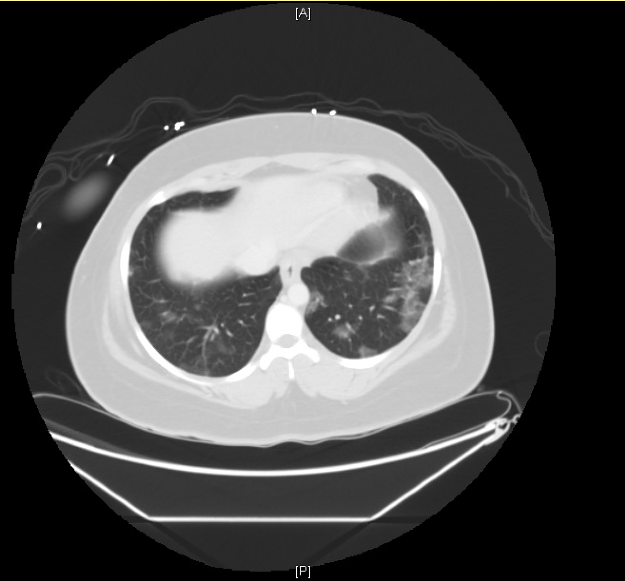
Cureus Ground Glass Opacities Observed In A 26 Year Old Coronavirus Disease 2019 Covid 19 Rule Out Patient With A History Of Vape Use

Tree In Bud Pattern In Central Lung Cancer Ct Findings And Pathologic Correlation Sciencedirect


